how do they x ray babies hips
A hip X-ray is a safe and painless test that uses a small amount of radiation to make images of the hip joints where the legs attach to the pelvis. I bawled my eyes out when they put him in it because he was so scared.

Uk Professor Says Swaddling Epidemic Gives Babies Clicky Hips Daily Mail Online Hips Professor Baby Swaddle
Asymmetrical buttock creases can suggest hip dysplasia in infants but like a hip click an ultrasound or x-ray study will need to be done to determine whether the hips are normal or not.

. The pics are hella. According to the makers website the Pigg-O-Stat is an all-in-one pediatric immobilization device designed for positioning infants and young children for an appropriate X-ray without significant. A hip X-ray can detect arthritis in the bones of that area.
During the examination an X-ray machine sends a beam of radiation through the pelvic bones and hip joints and an image is recorded on a computer or special film. This cast starts below the armpits and goes all the way. X-rays of the hip can reveal bone tumors and diagnose bone cancer.
The reported incidence of developmental dysplasia of the hip varies between 15 and 20 per 1000 births 1 with the majority 60-80 of abnormal hips resolving spontaneously within 2-8 weeks 1 so-called immature hip. On MRI the lesion is hyperintense on T2WI. The maximum visual information is given by the x-ray of the hip joint in two projections.
Appointments and Referrals. Youll be asked to partly undress your baby and take off their diaper for the test. X-rays of the bones taken over the years can show worsening of arthritis.
The American Academy of Pediatrics does not recommend routine ultrasounds for every infant. You will go in the room with him he will need to be stripped from the waist down they will take x-rays of him flat on his back legs dead straight and together you wil be able to hold him in this position then an x-ray of his still on his back with his knees bent facing outwards and the soles of his feet put together he will be fine its not traumatic at all you will be with him the. A few babies may need surgery to correct their hip joints.
Physicians use hip X-rays to locate foreign objects within the hip or that area of body and to guide them in setting broken bones. Hip Click Hip clicks or pops can sometimes suggest hip dysplasia but a snapping sound can occur in normal hips from developing ligaments in and around the hip joint. Its used because babies and toddlers are incapable of following directions to hold still.
Children use to grow and this means X-ray is done on baby bones and they will grow and growing tissue is altered by X-ray. An X-ray technician will take pictures of the hip. It might help to feed your baby just before the ultrasound to make your little one more relaxed.
You will go in the room with him he will need to be stripped from the waist down they will take x-rays of him flat on his back legs dead straight and together you wil be able to hold him in this position then an x-ray of his still on his back with his knees bent facing outwards and the soles of his feet put together he will be fine its not traumatic at all you will. Your baby will be in a spica cast for about 3 to 6 months which is upgraded as your baby grows. It has nothing to do with whether or not the parents will hold the infant.
A hip X - ray is a safe and painless test that uses a small amount of radiation to make images of the hip joints where the legs attach to the pelvis. The technologist will examine both hips of the child in different positions until. Babies must stay as still as they can during the examination so it is recommended that parents bring toys or soothe their babies with their voice to make time pass quicker.
Two tests are performed called the Barlow and Ortolani tests to examine the function of the hip joints. They should stay still for 23 seconds while each X-ray is taken so the images. An ultrasound may be needed to get a picture of the hip.
My 1st kid had 1 of these. F ratio 18 firstborn baby. While most people were laughing over the pictures some parents whose children had used the baby X-ray aka the Pigg O Stat said that the experience was anything but funny with some calling it traumatizing for their babies.
This means that babys thighs are spread around the parents torso and babys hips are open with his knees are bent at the same height as or higher than his bottom in an M shape like a sitting frog. They didnt warn me. Your baby will be placed on a table on their back or side.
How do they xray babies hips. Your baby will be put to sleep and the surgeon will set the ball joint in place. It works similar to a harness but its got a firmer hold and keeps their hips in place better.
In the direct projection or frontal obtained by focusing the x-ray tube perpendicular to the body plane - front or rear and axial transverse or horizontal plane fixing the elements of the joint from top to bottom - along the femur. A hip click can be felt by the examiner when the hip joints may not have formed normally. How do they xray babies hips.
Whichever style of baby carrier or sling you choose make sure your babys hips are spread out in the squat position. A Hip X-Ray may help diagnose find. The abscess is usually located in the metaphysis of long bones but may be located in the epiphysis in young children.
But for babies with an abnormal physical exam or major risk factors for developmental dysplasia of the hip or DDH family history Breech position etc the AAP supports referral for. Usually both hips are scanned for comparison. From the front anteroposterior view or AP from the side lateral view also known as the frog leg lateral view Typically X-rays of both hips are taken for comparison even if only one hip is causing symptoms.
Hip X-rays are done with a child lying on a table. The scan usually takes about 20 minutes. It depends on dose and tissue and the age of the patient.
In babies with hip dysplasia the joint has not formed normally and the hips are prone to moving in and out of joint. On x-ray there is a sharp defined oval lytic lesion with or without a sclerotic rim with its long axis parallel to the long axis of the bone see figure. During the examination an X-ray machine sends a beam of radiation through the pelvic bones and hip joints and an image is recorded on a computer or special film.
The question is the indication of X-ray on hip and legs. Because they spin around the body taking multiple images CT scans can deliver radiation doses that are up to 200 times higher than an average chest X. An X-ray of the pelvis focuses specifically on the area between your hips that holds many of your reproductive and digestive organs.
In addition exposing the parents to ionizing radiation X-rays needlessly goes against the ALARA principle. Risk factors include 14. A hip X-ray is a safe and painless test that uses a small amount of radiation to make images of the hip joints where the legs attach to the pelvis.

Pin On X Rays

X Ray Image Of Child Swallowed The Coins For A Medical Diagnosis Medicine Pictures Children Images X Ray Images

Basic Information About Dog Hip Dysplasia Paperblog Dog Hip Dysplasia Hip Dysplasia Canine Hip Dysplasia

One Month Post Op From A Right Periacetabular Osteotomy To Correct Hip Dysplasia Ehlers Danlos Syndrome Hip Dysplasia Surgery Recovery

Lower Limb Radiographs Anatomy And Physiology Anatomy Sacroiliac Joint

Congenital Hip Dislocation Chd Happens When A Child Is Born With An Unstable Hip Read On To Learn More Ab Canine Hip Dysplasia Hip Dysplasia Hip Dislocation

How To Shower After Hip Replacement Surgery Livestrong Com Hip Replacement Surgery Hip Replacement Exercises Hip Brace

Hip Dysplasia When You Re Too Young For A Hip Replacement Periacetabular Osteotomy Pao Bursitis Hip Hip Replacement Hip Replacement Surgery

My Hips Pre Pao Rpao January 2011 Rpao January 2011 Screws From My Rpao X Ray Ehlers Danlos Syndrome Surgery Recovery
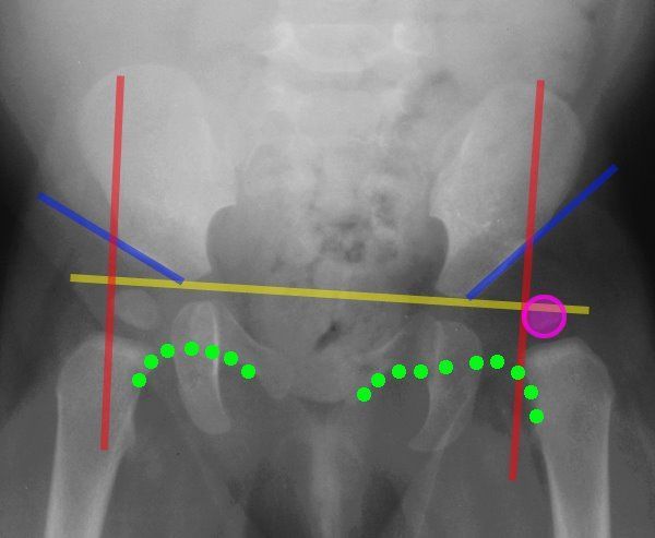
Lines Of The Hip Pediatrics Pediatrics Pediatric Nurse Practitioner Pediatric Radiology

Pin By Meg Carter On Ortho Hip Dysplasia X Ray Orthopedics

Pin On Teach

Pin On Adult Hip Dysplasia Awareness
Diagnosis Prevention And Management Of Canine Hip Dysplasia A Revie Vmrr Canine Hip Dysplasia Diagnostic Imaging Total Hip Replacement

Developmental Dysplasia Of The Hip Ddh Diagnostic Imaging Developmental Dysplasia Of The Hip Diagnostic Imaging Case Study

Severe Hip Dysplasia In A Boxer The Red Arrows Are Pointing To The Over Growth Of Bone At The Femoral Neck Head The Red Arrow Shades Of Grey Hip Dysplasia

Causes Of Ddh Hip Dysplasia Baby Developmental Dysplasia Of The Hip Baby Wearing

Hip Joint Developmental Hip Dysplasia 1 Year Old Child With A Dislocated Right Hip The Degree O Developmental Dysplasia Of The Hip Radiography Subluxation

Canine Ofa Hip Xrays Goldendoodle Puppy For Sale Labradoodle Goldendoodle Goldendoodle Puppy